AUTOMATED LIVE IMAGING OF HOST PATHOGEN INTERACTION FOR INNOVATIVE THERAPIES OF INFECTIOUS DISEASES (ALHOPATHI)
Coordinator :
Nathalie Aulner
Partners :
Financement :
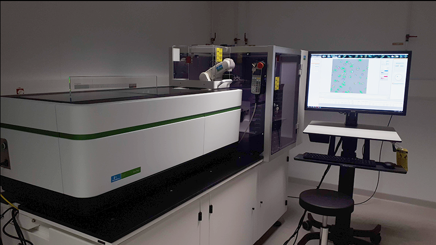
Opera Phenics
OBJECTIFS
The Institut Pasteur UtechS Photonic BioImaging facility plans to upgrade its BSL2+ high-content cell analysis and screening facilities with the installation of a latest generation automated spinning disk confocal cell imaging system. The system will be fully integrated into our level two access management biocontainment laboratory allowing a broad range of samples including human pathogens, and primary human cells to be safely handled in high-quality workflows. In this context our expert professional technology team will assure development and access to the state-of-the-art facilities benefiting Pasteurian researchers and the regional research community alike.
DESCRIPTION
Digital imaging provides a unique means to probe in situ functional and phenotypic biological information. Today, the cutting-edge of such methods combines Imaging with automated microfluidics, cytometry, and chemical-genomics. Here we propose to bring together the technology expertise and infrastructure capacity of scientific teams affiliated with the technology center Citech to hone new opportunities for experimental analyses. The current initiative will allow to bind multi-disciplinary expertise among Citech teams providing a full gamut of the necessary technologies for quantitative measure on the dynamics of cellular processes and their perturbation by disease, we will establish robust automated sample handling and data acquisition revealing quantitatively the interplay between host cell and pathogen, using immuno/chemical/genomic experimental perturbation. The state-of-the-art high-content cell imaging laboratory will offer sample manipulation, preparation and experimentation on virulent microorganisms. We will develop pertinent models using primary host cells, organ-on-a chip and iPS-derived cell based methods designed to mimic relevant physiological conditions. Ultimately this project will assure availability of world-class facilities for quantitative studies on clinically relevant models including viral infections, parasites, and bacteria.
MOYENS MIS EN OEUVRE
The requested equipment will be installed in the long-established Photonic BioImaging facilities (aka Imagopole) at the heart of the Citech technologies agglomeration of the Institut Pasteur. We will build upon a joint scientific and technology collaborative effort among the Citech facilities and scientific research teams to establish a “platform of know-how” for measuring dynamic cellular events occuring in disease processes. The imaging facility will provide access, training and support to the instrument, in a BSL2+ confinement equipped with a liquid-handling and pipetting robot. We will build upon our almost two decades experience in cell-based assay development to deliver the Pasteurian and french research community the full benefits of this investment (Aulner, Shorte et al.).

